Nociceptors
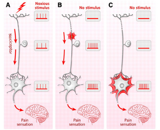
Figure 1: Nociceptor Detection
Nociceptors are the nerves within our body in charge of sending pain signals to the brain. They are what allow us to feel and sense external and internal pain infliction. Nociceptors also sense external temperature. They are located in skin, muscles and various organs.
The major types of pain detected by nociceptors are somatic pain and visceral pain. Somatic pain is pain that is generated from tissue damage, particularly deep tissue. A deep cut through the skin would be an example of a source of somatic pain. Visceral pain more so deals with pain generated within internal organs. Unlike somatic pain, visceral pain is more widespread and harder to detect the precise source. It can be described as more of an aching or squeezing type of pain.
Nociceptors work in a manner of detection and notification. They first detect the extreme temperature or pain stimulus and then that detection triggers the nociceptor to fire long range electrical signals through a pathway connected to the brain stem.
In Figure 1 part A, the process of nociceptor detection can be seen. Part B and C show a damaged nociceptor in various areas that can eventually lead to chronic pain.
The Pain and Temperature Pathway
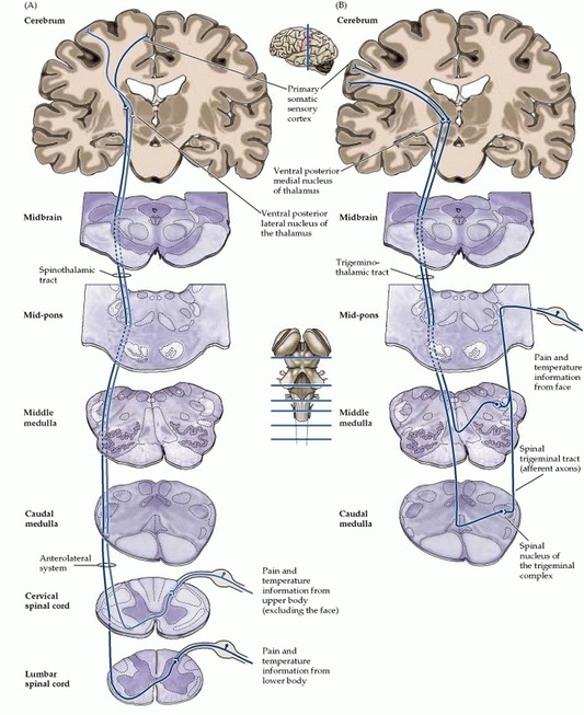
Figure 2: Pain and Temperature Pathway
Figure 2 provides a visual of the major pathways for pain and temperature sensation.
- Main pathways include the spinothalamic system and the trigeminal pain and temperature system.
- Axons of nociceptive nerve cells insert into the spinal cord through dorsal roots.
- Pain and temperature receptors are found however the more lateral area of the dorsal roots.
- Once in the spinal cord axons travel up and down what is called the dorsolateral tract of Lissauer.
- Once finished traveling through that region, the axons break through an area of the dorsal horn called “gray matter”.
- After leaving the dorsal horn, axons come in contact with neurons in the Rexed laminae I and II.
- Neurons will be transmitted to second order projection neurons that will finally reach the brainstem and eventually the thalamus.
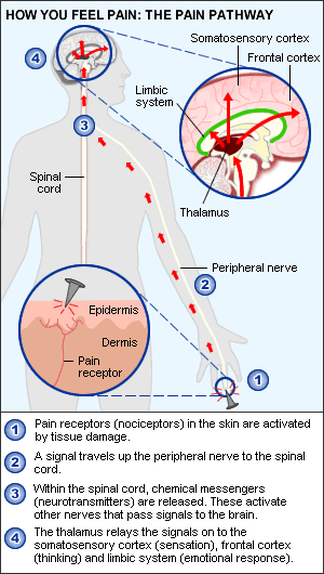
Figure 3: Pain Pathway
|
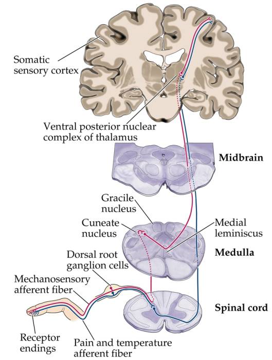
Figure 4: CNS Pain Pathway
|
As seen in figure 3, the pain is inflicted into the skin. The nociceptors are activated by this pain signal. The signal proceeds to travel up the peripheral nerve. The signal reaches the spinal cord and the brain stem. Once in the spinal cord, neurotransmitters are released activating various other nerves that sends signals to the stem of the brain. Once those signals reach the thalamus, the thalamus then relays those signals to the somatosensory cortex. This region of the brain deals with the body’s sensations. The signals are also relayed to the frontal cortex and limbic system which deal with thinking and emotional response respectively. This figure demonstrates the basic pathway when the skin or any part of the body endures extreme temperatures or pain.
Figure 4 depicts the pain pathway as well as the various area of the brain sensory pathways. Nociceptors originate from Dorsal root ganglion cells area. Pain nerve pathways are also connected to the spinal cord.
Figure 4 depicts the pain pathway as well as the various area of the brain sensory pathways. Nociceptors originate from Dorsal root ganglion cells area. Pain nerve pathways are also connected to the spinal cord.
Neurons
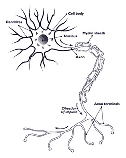
Figure 5: Neuron
Neurons are the functional units of the nervous system. [5] Together, different types of neurons carry signals over long distances to be relayed to the brain. A single neural cell has a general structure consisting of the cell body with dendrites, a long axon, followed by axon terminals. The dendrites of the neuron are responsible for receiving incoming signals from adjacent cells. The branch-like structure of dendrites increase the surface area of the cell body, thus allowing communication with multiple adjacent cells. The axon is the long body of the neuron, measuring up to a few micrometers to more than a meter, depending on neuron function. The axon is responsible for transmitting the received electrical signal to a neighboring neuron. On the end of the neuron, opposite to the dendrites, are the axon terminals, which secrete chemical or electrical signals to the dendrites of adjacent cells.
Two varieties of neurons are associated with pain signaling. Nociceptors are sensory neurons and can be found anywhere pain can be sensed. For example, nociceptors can be found on the skin, the cornea, and muscles, even though they are an external structure. Nociceptors are not found in the brain, which is why the brain itself does not feel pain directly. A bundle of neurons called the C nerve fibers are responsible for transmitting pain signals to the CNS. The neurons in C fibers have adjacent long axons, and are part of the peripheral nervous system. C fibers are not myelinated, making signal transduction slower compared to other myelinated neurons.
Two varieties of neurons are associated with pain signaling. Nociceptors are sensory neurons and can be found anywhere pain can be sensed. For example, nociceptors can be found on the skin, the cornea, and muscles, even though they are an external structure. Nociceptors are not found in the brain, which is why the brain itself does not feel pain directly. A bundle of neurons called the C nerve fibers are responsible for transmitting pain signals to the CNS. The neurons in C fibers have adjacent long axons, and are part of the peripheral nervous system. C fibers are not myelinated, making signal transduction slower compared to other myelinated neurons.
Membrane Potential of a Neuron
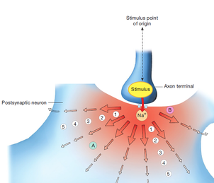
Figure 6: Graded Potential [5]
A single neuron, like all other living cells, has a characteristic membrane potential due to specific concentrations of Na+(sodium), K+(potassium), and Cl-(chloride) ions on both sides of the cell membrane. The Goldman-Hodgkin-Katz equation can be used to find the resting membrane potential of the neuron, given the concentrations of Na+, K+, and Cl- ions inside and outside of the cell membrane, as well as the permeability of each ion. The movement of an ion through an ion channel carries a current. Typically, K+ ions flow from inside the cell to outside the cell and Na+, Cl-, and Ca2+ flow from outside of the cell to inside the cell. The electrical signal from the neuron stems from the flow of current carried by ions entering or leaving the cell through ion channels. A neuron can become hyperpolarized or depolarized depending on the direction of ion flow.
Graded Potential
The electrical signal generated from hyperpolarization and depolarization of the neuron can be considered as an action potential or a graded potential. Graded potentials occur when ion channels are opened or closed in a neuron by reacting to chemical or mechanical stimuli from another neuron. [5] The effect of a graded potential is directly proportional to the amplitude of its electrical signal. An example of a graded stimulus can be seen in figure 6. The stimulus is delivered to a cell body of a neuron by the axon terminal of an adjacent cell. The stimulus chemically or mechanically opens the sodium gated ion channel, allowing the flow of Na+ ions to flow into the cell, depolarizing it. The strength of the overall potential depends on the initial contact with the stimuli. The stronger the stimuli, the greater the area of the neuron cell body will be affected. Figure 5 depicts that the strongest part of the signal, causing the most open sodium gated ion channels are found in areas labeled 1 or 2. Areas 3, 4, and 5 are further away from the stimulus, therefore have less open channels. In areas of the cell body further from the stimulus than area 5, the graded potential would have died out.
Action Potential
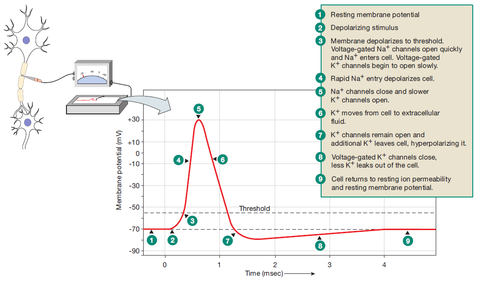
Figure 7: Action Potential [5]
An action potential occurs when a graded potential is strong enough to reach a trigger zone and cause a depolarization of about 100mV.[1] In a neuron, the trigger zone is located at the axon hillhock.[6] The strength of the graded potential does not matter, but for an action potential to occur, it must be able to depolarize the neuron to -55mV. Figure 6 illustrates the changes in membrane threshold potential during an action potential.
A neuron has a resting membrane potential of -70mV. At this stage 1, a resting potential of -70mV means that ion channels are closed. During stage 2, a graded potential would have reached the trigger zone and will have begun to depolarize the membrane to a threshold of -55mV. Stage 3 involves the opening of Na+ channels and the flow of Na+ ions into the cell. As a cation, the positive charge that Na+ carry continue to raise the membrane potential until about +30mV (Stage 4), to which then the sodium channels in the axon close, as depicted by stage 5. At this time in the action potential, the membrane polarity is reversed. Stage 6 of the action potential is due to the opening of voltage gated K+ channels in response to depolarization of the cell. Because of the positive potential, the opening of voltage gated K+ ion channels will cause K+ ions to flow out of the cell. The falling membrane potential is due to positive charge leaving the cell and eventually hyperpolarizing it. (Stage 7) At stage 8 and 9, Voltage-gated K+ channels would have closed and the cell returns to resting potential and ion permeability due to small leak channels that allow positive charge to flow.
A neuron has a resting membrane potential of -70mV. At this stage 1, a resting potential of -70mV means that ion channels are closed. During stage 2, a graded potential would have reached the trigger zone and will have begun to depolarize the membrane to a threshold of -55mV. Stage 3 involves the opening of Na+ channels and the flow of Na+ ions into the cell. As a cation, the positive charge that Na+ carry continue to raise the membrane potential until about +30mV (Stage 4), to which then the sodium channels in the axon close, as depicted by stage 5. At this time in the action potential, the membrane polarity is reversed. Stage 6 of the action potential is due to the opening of voltage gated K+ channels in response to depolarization of the cell. Because of the positive potential, the opening of voltage gated K+ ion channels will cause K+ ions to flow out of the cell. The falling membrane potential is due to positive charge leaving the cell and eventually hyperpolarizing it. (Stage 7) At stage 8 and 9, Voltage-gated K+ channels would have closed and the cell returns to resting potential and ion permeability due to small leak channels that allow positive charge to flow.
References
- http://www.ncbi.nlm.nih.gov/pmc/articles/PMC2964977/
- http://pain.about.com/od/typesofchronicpain/g/nociceptors.htm
- http://www.ncbi.nlm.nih.gov/books/NBK10965/
- www.nutritionreview.org
- Silverthorn, DU. Human Physiology. San Francisco: Pearson. p 249-263.
- Lodish H, Berk A, Zipursky SL, et al. Molecular Cell Biology. 4th edition. New York: W. H. Freeman; 2000. Section 21.1, Overview of Neuron Structure and Function. Available from: http://www.ncbi.nlm.nih.gov/books/NBK21535/
